
INTRODUCTION & SCOPE
1. Medical Terminology – Define or Explain:
a. Infection: “Entry” into a
host of “Infectious agent”
f. Infectious
Agent: ( There are “3”)
Pathogens, Non-pathogens and Normal Body Flora
Pathogens, Non-pathogens and Normal Body Flora
infectious agent: Medical term for
germ, parasites
Infectious
agent that is harmful – Pathogen
Pathogen- “disease-producing”
infectious agent
Infectious agent that causes harm –
Disease
Host with Disease – Patient
Infectious agent that is not harmful – non-pathogen
b. Invasion-
is an infectious agent that:
Settles into the infected host
Establishes itself
Multiplies and spreads in the host
after infection
“Takes over after infecting the host”
g. Normal Body Flora:Germs
that establish a fairly (more or less) permanent
residence, but do not cause disease under normal conditions.
residence, but do not cause disease under normal conditions.
They are members
of the bodys Noramal microbiota or Normal
flora
flora
Example: Lactobacilli- the first
microorganisum that enters a
newborn
newborn
E. coli – is generally harmless (non-pathogen) but if it
crossed into other parts, it becomes Pathogens
crossed into other parts, it becomes Pathogens
c. Etiology- The study
of “cause” of disease
h. Disease – When
infection results in change of state of health
2. Demonstrate Understanding of:
a. Pathology- is the ”study”
of disease
d. Pathogens – (Harmful)
infectious agent that causes disease / suffering
Non-Pathogens – (Harmless)
b. Pathogenicity –
Clinical term for the “ability” of germs to cause disease
e. Opportunistic Pathogen
– Are
ususally non-pathogens (harmless) but can become
Harmful, depending on the environment or host. When
the host becomes weak or substance change, they
become pathogens :
Harmful, depending on the environment or host. When
the host becomes weak or substance change, they
become pathogens :
They are: Disease,
Harmless (depending on condition of the host)
Neutral
Noramal Body Flora (depending on environment)
example:
E coli ( Harmless in intestine) Harmful in other
parts
parts
c. ‘HAI’ –“Healthcare associated infection”
15% of patients can become victims
- to avoid this …….
“Chain of transmission” should be controlled or
avoided using scrubs,
cloth ect outside of authorized areas.
cloth ect outside of authorized areas.
f. Nosocomial Infections –
Infection as a result of “hospital stay”
(microorganisms in hospital)
(microorganisms in hospital)
8th leading cause
of death in the USA
3. Spontaneous
Generation, Biogenesis, Germs and Disease
a. Recall Scientists who supported Spontaneous Generation :
- ” John
Needham”
c. Identify Scientists associated with Microscopy
1. Robert Hooke- Invented
the “compound microscope”
Invented the lens and condenser, the condenser focuses
light on the sample with a bottle of water.
light on the sample with a bottle of water.
2. Antonie van Leeuwenhoek: made
“simple microscope”, with 1 lens
Observed first
bacteria, in his drawings:“the father of microbiology”
3. Louis Joblot- added
the “side pillar and iris diaphragm”
Iris diaphragm- (is the light adjuster).
Side pillar- (is the arm)
b. Learn about "Germ
Theory of Disease" and its Scientists:
The Germ Theory of Disease discovered by:
Louis Pasteur
He
determined that “germs cause disease”
Microbes are in
the air
d. Study Koch's Postulates
and the five key steps:
Koch is “the father of
etiology”
Linked “specific
microorganisms to specific disease”
Anthrax: is caused by Bacillus
anthraxis
Koch's Postulates and the “
five key steps” :
2. “Pathogen must be isolated”
(Take sample of pathogen) from
sick host & grown in pure culture.
sick host & grown in pure culture.
3. “Inoculated (inject) it
into a healthy, susceptible host- (Host #2) “ (duplicate
the disease)
4. Pathogen isolated in
culture must produce similar symptoms; (Now it
reproduced in Host #2)
5. Pathogen
from new Host (Host #2) must match profile of original culture:
e. Study properties of Normal Body Flora
4. Systems of Measurement
4. Systems of Measurement
Compare Imperial and Metric System for:
a. Size
c. Temperature
Length
| Metric | US or Imperial | ||
|---|---|---|---|
| 1 millimeter [mm] | 0.03937 in | ||
| 1 centimeter [cm] | 10 mm | 0.3937 in | |
| 1 meter [m] | 100 cm | 1.0936 yd | |
| 1 kilometer [km] | 1000 m | 0.6214 mile | |
| US or Imperial | Metric | ||
|---|---|---|---|
| 1 inch [in] | 2.54 cm | ||
| 1 foot [ft] | 12 in | 0.3048 m | |
| 1 yard [yd] | 3 ft | 0.9144 m | |
| 1 mile | 1760 yd | 1.6093 km | |
| 1 int nautical mile | 2025.4 yd | 1.853 km | |
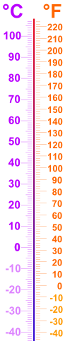
c. Temperature
Conversion of Temperature
Quick Celsius (°C) / Fahrenheit (°F) Conversion:
Formula to convert F to C or C to F F=( 1.8 x C) +32
Typical Temperatures
|
 |
b. Weight
d. Volume
Mass
| Metric | US or Imperial | ||
|---|---|---|---|
| 1 milligram [mg] | 0.0154 grain | ||
| 1 gram [g] | 1,000 mg | 0.0353 oz | |
| 1 kilogram [kg] | 1,000 g | 2.2046 lb | |
| 1 tonne [t] | 1,000 kg | 1.1023 short ton | |
| 1 tonne [t] | 1,000 kg | 0.9842 long ton | |
| US or Imperial | Metric | ||
|---|---|---|---|
| 1 ounce [oz] | 437.5 grain | 28.35 g | |
| 1 pound [lb] | 16 oz | 0.4536 kg | |
| 1 stone | 14 lb | 6.3503 kg | |
| 1 hundredweight [cwt] | 112 lb | 50.802 kg | |
| 1 short ton (US) | 0.9072 t | ||
| 1 long ton (UK) | 1.0160 t | ||
d. Volume
Volume/Capacity
| Metric | US Measure | Imperial | ||
|---|---|---|---|---|
| 1 cu cm [cm3] | 0.0610 in3 | |||
| 1 cu decimeter [dm3] | 1,000 cm3 | 0.0353 ft3 | ||
| 1 cu meter [m3] | 1,000 dm3 | 1.3080 yd3 | ||
| 1 liter [l] | 1 dm3 | 2.113 fluid pt | 1.7598 pt | |
| US Measure | Imperial | Metric | |
|---|---|---|---|
| 1 cu inch [in3] | 16.387 cm3 | ||
| 1 cu foot [ft3] | 0.02832 m3 | ||
| 1 fluid ounce | 1.0408 UK fl oz | 29.574 ml | |
| 1 pint (16 fl oz) | 0.8327 UK pt | 0.4732 liters | |
| 1 gallon (231 in3) | 0.8327 UK gal | 3.7854 liters | |
5. Units and Measurements:
-When measuring microorganisms - metric system is used, the standard unit of length in the metric system is the meter (m)
-major advantage is - all units are factors of 10
-to reduce- you reduce by factors of 10
-cells are measured in - micro
- Viruses - are measured by nano
- viruses are smaller then bacteria
Determine the relative
values and symbols for these sub-units:
a. Deci- 1 decimeter = .1 or 1/10th of base unit
d. Micro-1 micrometer =1/1,000,000. (1 in a million)
d. Micro-1 micrometer =1/1,000,000. (1 in a million)
b. Centi- 1 centimeter = .01
e. Nano- 1/Billion (1 in a billion)
e. Nano- 1/Billion (1 in a billion)
c. Milli-.001 or 1/1000 -
f. Pico- 1/1,000,000,000,000 (1 in a trillion)

f. Pico- 1/1,000,000,000,000 (1 in a trillion)
6. Microscopy
a. Compare Simple and Compound Microscopes
a. Compare Simple and Compound Microscopes
simple microscope- (Antoni van Leeuwenhoek)
- 0ne lens
- Location of specimen on pin (stage)
- Focusing control (the adjustment knob)
- Stage - positioning screw
Compound microscope-
- Lens and condenser
- has multiple lenses that work together so that lens #2 must
start where lens #1 left off.
- Tense to be Binocular
- Tense to be light (now days we use ligh bulbs)
-Now days we have "Binocular compound light Microscopes "
- monocular microscope- has only One eye piece and regular(4)
objective lenses..
-Binocular microscope- has Two eye pieces and regular
(4)objective lenses
b. Name Microscope Parts and identify their functions
page 55 ( - magnificarion is achieved by )
light rays from an illuminator (light source) passes thru a condendser (focuses light thru specimen) then light passes into the objective lense (primary lens that magnifies the specimen - lens #1) and the image of the specimen get magnified again by the ocular lens or eye piece (re-magnifies the image formed by the objective lens - lens #2)
Other parts:
- Body Tube -connects lens #1(objective lense) to lens #2(Ocular Lens)
- Arm
-Stage - Holds the microscope slide in position
-Diaphragm - Controls the amount of light entering the condenser
- Course focusing knob - The big knob ????
- Fine focusing knob - small knob ????
- Chapter title: Microscopes
- A list of vocabulary words is found toward the end of this document $ lecture: Microscopes
- Scale of microorganisms
- Micrometer (�m) = 10-6 meters <= bacteria cell (0.1 �m ~ viruses); nanometer (nm) = 10-9 meters ~ an amino acid; angstrom (Å) = 10-10 meters ~ an atom.
- Microscope
- a device used to make enlarged images of minute objects.
- Light microscopy
- A microscope which uses visible light to form the observed enlarged image. Contrast with electron microscopy.
- Note that light microscopes are unable to visualize viruses.
- Simple microscope
- A very minimal light microscope having only a single lens.
- Compound microscope
- A light microscope consisting of multiple lenses.
- A contemporary compound microscope is capable of magnification of up to 2000x and resolution of up to 0.2 �m.
- Critical parts of a compound microscope include
- the ocular
- the objective lenses
- the stage
- the condenser
- the diaphragm
- the illuminator
- one or more focusing knobs.
- See fig 3.1, p. 54, Tortora et al., 1995.
- Magnification
- The perceived increase in size of an object upon viewing through a microscope.
- Total magnification obtained with a compound microscope is equal to the product of the magnification supplied by the ocular and that supplied by the objective lenses (e.g., 10X * 100X = 1000X).
- Note that for all but the best light microscopes, a magnification of 1000X is the best that can be achieved.
- Resolution [resolving
power]
- The ability of a microscope to present a magnified image of two closely spaced points as separate points.
- A microscope with poor resolving power may present an image in which two closely spaced points do not appear as two separate images.
- The resolution of a microscope is measured in units of distance between these two hypothetical points.
- A microscope is only as useful as it is able to resolve the structures you are interested in viewing.
- Ocular
- The last lens or eyepiece of a compound microscope.
- The ocular imparts the final magnification of the image observed.
- It often magnifies on the order of 10X.
- Objective lens
- The first lens of a compound microscope.
- This is the lens closest to the specimen being observed.
- Different objective lenses are used in order to impart difference degrees of magnification.
- Magnification imparted by the objective lens often ranges from 10X, to 40X, to 100X or even 200X (the two latter magnifications achieved with oil immersion).
- Focal length
- Engraved on the side of objective lenses is both the power of the lens (e.g., 10x) and the focal length (e.g., 16 mm).
- Note that the focal length indicates how close the objective lens being used should be to the sample in order to bring the sample into sharp focus.
- Stage
- The often movable platform which holds the specimen.
- Condenser
- Lenses found after the illuminator but prior to the specimen.
- The condenser focuses the light passing through the specimen and into the objective lens.
- Diaphragm
- Adjusts to control the amount of light coming from the illuminator which enters the rest of the microscope through the condenser.
- Illuminator
- The light source for the compound microscope.
- This is usually an incandescent light bulb which is located at the bottom of the compound microscope.
- Focusing knobs
- These knobs move the stage up and down so that the relevant horizontal plane in the specimen is brought into focus.
- Often a compound microscope will have both a coarse and a fine focusing knob.
- Refraction [refraction
index]
- Deflection of light from a straight line occurring upon passing from one substance to another through a slanting interface (so long as the refractive indices of the two substances are different).
- Refraction causes a loss of light and therefore darkening.
- Such darkening is seen as images using the compound microscope.
- Staining and other means (e.g., phase-contrast microscopy) are employed to increase the refraction displayed by specimens and therefore their visibility.
- Oil immersion
- A technique employed to increase resolution at higher magnifications.
- Oil with the same refractive index as the glass slide holding the specimen is placed between the specimen and the objective lens.
- This prevents light from refracting between the specimen and the objective lens and images are consequently sharper.
- Brightfield microscopy
- Use of a compound microscope such that the image observed is the product of light that has passed through the specimen.
- It is called brightfield because where there is no specimen the light observed is brightest.
- Thus, the background is brighter than the specimen.
- Because brightfield microscopy relies solely on refraction, most specimens are difficult to see without staining.
- See fig 3.4a, p. 56, Tortora et al., 1995.
- Darkfield microscopy
- In darkfield microscopy there is an opaque disk in the condenser which blocks all light passing in a straight line from the condenser, through the specimen, into the objective lens.
- As a consequence, only scattered and reflected light is observed and the background is dark.
- This is one way of observing microorganisms without staining and therefore is useful for observing bacteria which are not easily stained.
- It necessarily gives a different image from that seen when employing brightfield microscopy and staining.
- Darkfield microscopy is somewhat archaic since today more sophisticated methods are available for visualize stain-resistant bacteria.
- Phase-contrast microscopy
- A way of viewing free, unfixed microorganisms with good detail.
- Phase-contrast microscopy relies on the differential diffraction (caused by refraction) of the light passing through the specimen, its different component parts, and the suspending medium.
- The instrument is adjusted such that all light passing through the medium (background) remains in phase and light passing through the specimen does not thus resulting in a specimen that is darker than the background.
- As better put by Burrells, 1977: "A phase microscope is a devise which causes difference of refractive index between an object and its surrounding medium to be made visible in the form of an ordinary black and white image.
- The term �phase contrast� has come to be used because the differences in phase of some rays in the light bundle passing through the system are used to give the necessary �contrast� between the objective and the background."
- Electron microscopy (EM)
- A microscope employing electrons instead of light to image specimens. Electron microscopy, or EM, is capable of much better resolution than light microscopes because the wavelength of electrons is much smaller than that of the photons of visible light.
- Transmission electron
microscopy (TEM)
- A type of EM used to examine very thin sections of fixed, stained, and dehydrated specimens.
- Gives magnifications ranging from 10,000X to 100,000X.
- Scanning electron microscopy
(SEM)
- A type of EM used to examine the surfaces of intact specimens.
- You�ve all seen what the 3-D-looking SEM images look like, such as photographs that appear to be extreme close-ups of insects.
- SEM is capable of magnifying in the range of 1000X to 10,000X and resolving objects as close together as 20 nm.
- Staining with biologic dyes
- The coloring of microorganisms with dyes (chromophoric ions) that bind specifically to various cell structures.
- Fixation
- The binding of microorganisms to the microscope slide. This prevents them from being washed off the slide during staining. Fixation is usually accomplished by spreading (smearing) the organism onto the slide, drying, and then flaming the slide lightly. Fixation usually results in the death of the attached microorganisms.
- Basic dyes
- A stain that binds to acidic substances such as bacteria.
- Acidic dyes
- A stain that binds to basic substances. These tend to be the background and acidic dyes thus tend to give negative staining.
- Negative staining
- A staining method whereby the specimen is left unstained but visible against a stained background.
- Simple stain
- A stain employed to highlight the entire microorganism (as opposed to differentiating its component parts) using only a single type of dye.
- Contrasts with differential staining.
- Mordant
- A substance used to increase the ability of a stain to highlight the microorganism or its component parts.
- Differential stains
- A staining method employed to distinguish among different kinds of bacteria.
- That is, different types of bacteria are stained differently.
- Differential stains are not simple because at least two stains are necessary to differentiate two organisms while simultaneously assuring that both organisms are stained by at least some dye.
- Gram stain
- A method of differentiating bacteria by staining and brightfield microscopy into two broad, phylogenetically relevant groups: gram-negative bacteria and gram-positive bacteria.
- Gram stains are performed as follows:
- Bacteria are heat-fixed to a slide.
- A purple basic dye such as crystal violet is applied to the fixed smear as the primary stain.
- After a short period the primary stain is washed off with distilled water. The cells are now stained purple.
- The washed fixed smear is then covered with an iodine mordant.
- The iodine is then washed off with distilled water. The cells are now stained darker purple.
- The washed fixed smear is then decolorized by further washing with either ethanol or a solution consisting of ethanol and acetone. This washing removes the purple color (primary stain) from gram-negative bacteria but not from gram-positive bacteria.
- The decolorizing agent is washed off with distilled water.
- The decolorized and washed fixed smear is then counterstained with safranin, a red basic dye.
- The safranin stained fixed smear is again washed with distilled water. Gram-positive bacteria retain the primary stain as above and remain purple but gram-negative bacteria do not retain the primary stain following decolorizing and are consequently stained only by the counterstain. Gram-negative bacteria are thus stained red following safranin counterstaining.
- The slide is blotted dry and examined by brightfield microscopy. Note that gram staining is best done using growing (exponential phase) bacteria and that some bacteria are resistant to staining and thus cannot be successfully gram typed.
- Primary stain
- A simple stain employed in the course of differential staining.
- The function of the primary stain is to impart a color to the cell present regardless of their potential for accepting the differential stain.
- Counterstains
- A stain with a contrasting color to a primary stain.
- Acid-fast stain
- A method of differential staining that detects the presence of waxy cell wall material.
- Acid-fast staining is useful for bacteria which are not readily gram stained and particularly for identifying Mycobacterium spp.
- Special stains
- Stains used to view specific cellular structures such as capsules, endospores, and flagella.
- Vocabulary
- Acid-fast stain
- Acidic dye
- Basic dye
- Brightfield microscopy
- Compound microscope
- Condenser
- Darkfield microscopy
- Diaphragm
- Differential staining
- Fixation
- Focusing knobs
- Gram stain
- Illuminator
- Mordant
- Negative staining
- Objective lens
- Ocular
- Oil immersion
- Phase-contrast microscopy
- Primary stain
- Resolution
- Resolving power
- Scanning electron microscopy (SEM)
- Simple stain
- Stage
- Transmission electron microscopy (TEM)
- Practice questions
- Using a good compound light microscope with a resolving power of 0.3 �m, a 10X ocular lens, and a 100x oil immersion lens, would you be able to discern two objects separated by 3 �m? 0.3 �m? 300 nm? 3000 Å? [PEEK]
- Acidic dyes stain the __________ in a smear and are used for ___________ staining (fill in the blanks and don�t use the term simple for the second blank). [PEEK]
- What is the purpose of the counterstain in staining techniques such as the gram stain? [PEEK]
- Name a reason for using phase contrast microscopy rather than brightfield microscopy ? [PEEK]
- Describe why in gram staining only gram-positive cells retain the primary stain? [PEEK]
- Practice question answers
- You would be able to discern two objects separated by the four distances given because each is equal to or greater than the resolving power of the microscope.
- In this order the blanks should be filled with: (i) background, (ii) negative.
- to visualize otherwise unstained microorganisms
- Viewing living, unfixed, unstained cells.
- The key word is retain. The primary stain is retained by gram-positives because it makes an intracellular complex with the iodine mordant that stays in place so long as the cell envelope is reasonably intact. The decolorizing agent breaks down the cell envelope of the gram-negative (but not gram positive) cells sufficiently that the primary stain-mordant complex is lost (washed out). The difference between cell types is that the gram negative cell envelope is thin and includes the outer membrane, the latter which is particular susceptible to the decolorizing solvents. The answer: The primary stain-mordant complex is retained in the gram-positive cell but not gram negative cells because of differences in cell envelopes, was sufficient.
- References
7. Principles of Microscopy
a. Identify the lenses of
a Compound Microscope:
Objective lens and Ocular lens
Objective lens and Ocular lens
b. Name, list magnifying power, and color codes to Objective lenses.
- Scanning - (4x) objective - red
- Low Power - (10x) objective - yellow
- High Power - (40X) Objective - Blue
- Oil Immersion - (100x) objective - White (Must have Oil)
c. Learn How to calculate
Total Magnification
- Scanning - (4x) = 40
- Low Power - (10x) = 100
- High Power - (40X) = 400
- Oil Immersion - (100x) = 1000
Example:
Objective lens power + Ocular Lens Power = Total Magnification
- Scanning - (4x) = 40
- Low Power - (10x) = 100
- High Power - (40X) = 400
- Oil Immersion - (100x) = 1000
Example:
Ocular lens magnification = 10X
Objective lens magnification being used = 4X
Total magnification? Calculation: (10X)(4X) = 40X
Objective lens power + Ocular Lens Power = Total Magnification
8. Resolution
a. Define Resolution, Useful, Empty Magnification
Resolution: is about seeing very fine details l!
( also called resolving power)
- its the ability for lens to distinguish fine details and
structures. Specifically, the ability of the lens to
distinguish two points a specific distance apart.
- Determines- "minimum spacing" or needed to see two
objects(adjacent) separately.
-Determines- the smallest size an eye can see
- the limit for the human eye to see is
200 (micrmeters), SEM200 (example $5 bill details),
less then 200 (micrometers) (300,400, 500 ect)
you can not see them ,too
small.
- ("Magnification can potentially improve resolution")
-(Two) aspects of resolution is .....
Useful magnification and Empty Magnification
Useful Magnification: ( everything we did with bills is useful magnification)
- "Increasing" magnification the reveals
"NEW" DETAILS and INFORMATION" because a
"HIGHER" resolution was produced.
- (nose-piece) - was introduced to improve
magnification (multiple objective lenses with
different / higher power options
- Its usually has 4 lenses...
- Useful magnification drove the invention of the
nose-piece.
Empty Magnification - "increasing magnification "does not" release
new details because the resolution has not
changed. example a TV
- Since the image is large, its easier to see and you
don't want to see details.
- Lens - High Power 40X and Oil immersion 100X or empty
magnification
b. Determine Resolution and/or Total Magnification for:
i. the Natural eye 200 micrometers
ii. 4x Objective lens 40
iii. 10x Objective lens 100
iv. 40x Objective lens 400
v. 100x Objective lens 1000
c. How does Resolution vary with:
c. How does Resolution vary with:
i. Lens Aperture Size-
Aperture Size - is the size of the opening on the
odjective lens
- resolution is "Higher" with "New Details" as
magnification "increases" the aperture
size "decreases".
- You can link aperture size to magnification
Aperture Size - is the size of the opening on the
odjective lens
- resolution is "Higher" with "New Details" as
magnification "increases" the aperture
size "decreases".
- You can link aperture size to magnification
ii. Working Distance -(the distance between Objective lens and slide)
- as magnification "Increases" the working
distance "decreases" wheras, the numerical
aperture of the objective lens increases. (lab pg 8)
The working distance for lens to work properly:
4X - 9-10mm
10X - 5- 8mm
40X - .5 - .7mm
100X - .13 - .18mm
Distortion - 130 - 180mm
- as magnification "Increases" the working
distance "decreases" wheras, the numerical
aperture of the objective lens increases. (lab pg 8)
The working distance for lens to work properly:
4X - 9-10mm
10X - 5- 8mm
40X - .5 - .7mm
100X - .13 - .18mm
Distortion - 130 - 180mm
iii. Body tube length - (Robert Hooke introduced the the body tube)
- a longer tube "increases magnification" with a "potential"
for higher resolution....
- How far should the Body tube length and objective lens be?-
(160mm / 16cm for all 4 lenses.)
9. Refraction and Refractive Index
a. What is refraction? the light bending ability of transparent
materials which may cause distortion of the "images" they
form.
- Your Slide is a potential place for refraction
Slides are very thin so light can hurry thru without refraction
your light goes from stage to specimen to air (working distance) part of the image is in the refraction. 4x has huge aperture size so the image will stay in focus and bring everything to range.
b. What is effect of
refraction of image formation?
-
-
c. Recall Refractive
Indexes for common items listed in class
d. How does Immersion oil
correct refraction?
- without oil , most light will refracted and lost.
- Benefits of oil - Eliminating air from working distance.
- essentially "recover refracted light"
- and reduces or minimizes refraction by squeeze it into aperture
size.
Oil is equivalent to liquid glass in working distance
- the image becomes sharply focused and better resolution.
(four principle Advances in microscopes.)....
Resolution
illuination
contrast
image quality
10. Types of Compound Light Microscopes
Describe Compound Light
Microscopes and Compare them
to highlight differences
in design and image quality for:
a. Brightfield - Dark objects are visible against a
bright background
- you DIE organism for contrast
bright background
- you DIE organism for contrast
b. Darkfield - Dark night effect
- Has "OPAQUE DISC" - this blocks light from
entering directly
- used to examine live microorganisms that are
invisible in ordinary light or can not be stained
by standard method or too distorted and cant be
identidied pg 57
entering directly
- used to examine live microorganisms that are
invisible in ordinary light or can not be stained
by standard method or too distorted and cant be
identidied pg 57
No comments:
Post a Comment